43 human eye with labels
FREE! - The Human Eye Labeling Activity (Teacher-Made) In this resource, you'll find a 2-page PDF that is easy to download, print out, and use immediately with your class. The first page is a labelling exercise with two diagrams of the human eye. One is a view from the outside, and the other is a more detailed cross-section. On the second page, you'll find a set of answers showing the properly labelled human eyes, designed to help you check ... Human Eye Anatomy Stock Photos, Pictures & Royalty-Free ... Anatomy of human eye and descriptions. Parts of the eye, labeled vector illustration diagram How eye work medical illustration, eye - brain diagram, eye... human blue eye extreme macro Eye anatomy. Rod cells and cone cells. Anatomy of human eye hand draw vintage clip art isolated on... eyeball Human eye anatomy Human eye anatomy vector design
The Eyes (Human Anatomy): Diagram, Optic Nerve, Iris ... Eye Conditions. Age-related macular degeneration: Causes loss of central vision as you get older.. Amblyopia: Often called lazy eye, this condition starts in childhood.One eye sees better than the ...

Human eye with labels
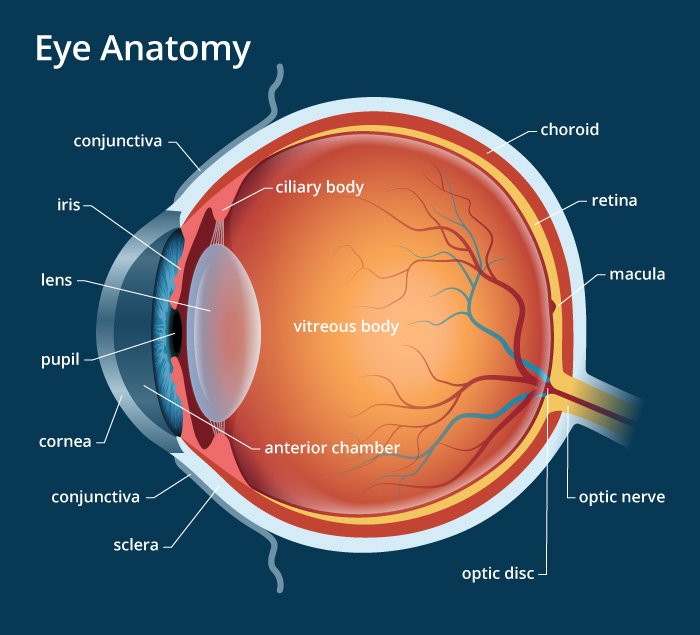
Human eye - Wikipedia The human eye is a sensory organ, part of the sensory nervous system, that reacts to visible light and allows us to use visual information for various purposes including seeing things, keeping our balance, and maintaining circadian rhythm . The eye can be considered as a living optical device. Eye Diagram - Labelled Diagram of Human Eye, Explanation ... The human eye is a part of the sensory nervous system. Labeled Diagram of Human Eye The eyes of all mammals consist of a non-image-forming photosensitive ganglion within the retina which receives light, adjusts the dimensions of the pupil, regulates the availability of melatonin hormones, and also entertains the body clock. Labeled Eye Diagram | Eye anatomy diagram, Eye anatomy ... This Article is the detailed account of all the major organs that are categorized under the nine regions in the abdominal cavity 1) Stomach 2) Intestines a) Small Intestine Duodenum Jejunum Ileum b) Large Intestine Ceacum Colon (Ascending, Transverse and Descending) Rectum Anal Canal 3) Liver 4) Gall bladder 5) Pancreas 6) Spleen 7) Kidneys […] S
Human eye with labels. Eye Diagram - Differentiated Worksheets and ... - Pinterest Jan 29, 2016 - Use these simple eye diagrams to help students learn about the human eye. Three differentiated worksheets are included: 1. Write the words using a word bank2. Cut and paste the words3. Write the words without a word bank Labels include: eyebrow, eyelid, eyelashes, pupil, iris, and sclera.UPDATE:I'... Labeled Eye Diagram - Science Trends What you want to interpret as a major part of the human eye is somewhat up to the individual, but in general there are seven parts of the human eye: the cornea, the pupil, the iris, the lens, the vitreous humor, the retina, and the sclera. Let's take a closer look at each of these components individually. The Cornea Labelling the eye - Science Learning Hub In this interactive, you can label parts of the human eye. Use your mouse or finger to hover over a box to highlight the part to be named. Drag and drop the text labels onto the boxes next to the eye diagram If you want to redo an answer, click on the box and the answer will go back to the top so you can move it to another box. Anatomy of the eye: Quizzes and diagrams | Kenhub Try our crash course in eye anatomy. One of our favorite ways to get to grips with all of the parts of the eye is by utilizing labeled diagrams. On a diagram of the eye, we can see all of the relevant structures together on one image. This helps us to understand how each one is situated and related to the other. Labeled diagram of the eye
FREE! - Label the Eye Worksheet - Teacher-Made Learning ... In this resource, you'll find a 2-page PDF that is easy to download, print out, and use immediately with your class. The first page is a labelling exercise with two diagrams of the human eye. One is a view from the outside, and the other is a more detailed cross-section. On the second page, you'll find a set of answers showing the properly labelled human eyes, designed to help you check ... Human eye diagram Images, Stock Photos & Vectors ... Human eye diagram images 6,714 human eye diagram stock photos, vectors, and illustrations are available royalty-free. See human eye diagram stock video clips Image type Orientation Sort by Popular Biology Healthcare and Medical Diseases, Viruses, and Disorders human eye anatomy eye retina medicine visual perception cone cell Next of 68 Eye Anatomy: Parts of the Eye and How We See - American ... The eye sits in a protective bony socket called the orbit. Six extraocular muscles in the orbit are attached to the eye. These muscles move the eye up and down, side to side, and rotate the eye. The extraocular muscles are attached to the white part of the eye called the sclera. This is a strong layer of tissue that covers nearly the entire ... PDF Eye Anatomy Handout - National Eye Institute of light entering the eye. Lens: The lens is a clear part of the eye behind the iris that helps to focus light, or an image, on the retina. Macula: The macula is the small, sensitive area of the retina that gives central vision. It is located in the center of the retina. Optic nerve: The optic nerve is the largest sensory nerve of the eye.
Human Eye Diagram, How The Eye Work -15 Amazing Facts of Eye Fun Facts About Human Eye For Kids FACT 1 Iris scanning is more secure than fingerprints because our iris has 256 unique characteristics and the fingerprint has just 40. FACT 2 Newborn babies don't produce tears. They only make crying sounds, but no tears come out of their crying eyes. Structure and Functions of Human Eye with labelled Diagram The human eyes are the most complicated sense organs in the human body. From the muscles and tissues to nerves and blood vessels, every part of the human eye is responsible for a certain action. Furthermore, contrary to popular belief, the eye is not perfectly spherical; instead, it is two separate segments fused together. 43 diagram of the human eye without labels Label Parts of the Human Eye - University of Dayton Label Parts of the Human Eye. Select One Anterior Chamber Ciliary Body Cornea Fibrous Tunic Iris Lateral Rectus Muscle Lens Medial Rectus Muscle Optic Disk Optic Nerve Pupil Retina Vascular Tunic Vitreous Nerve. Human Eye Anatomy - Parts of the Eye ... Label Parts of the Human Eye - University of Dayton Parts of the Eye Select the correct label for each part of the eye. The image is taken from above the left eye. Click on the Score button to see how you did. Incorrect answers will be marked in red.
Eye Colors: Iridology Iris Pictures
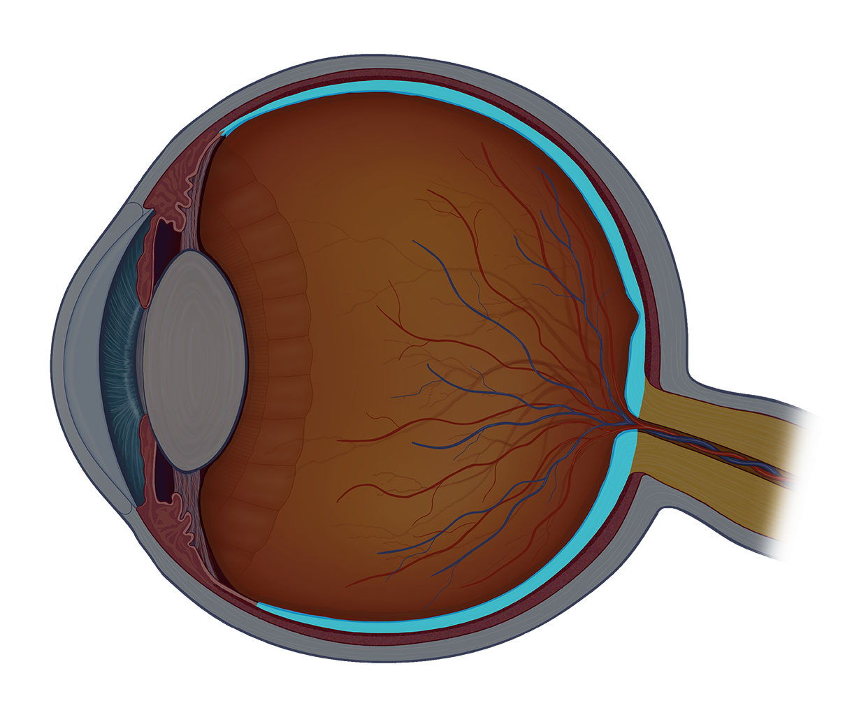
File:Diagram of human eye without labels.svg - Wikimedia ... File:Diagram of human eye without labels.svg. Size of this PNG preview of this SVG file: 410 × 430 pixels. Other resolutions: 229 × 240 pixels | 458 × 480 pixels | 732 × 768 pixels | 976 × 1,024 pixels | 1,953 × 2,048 pixels.

Human Anatomy Lab: The Respiratory System
Label Eye Printout - EnchantedLearning.com Label the Eye Diagram. Human Anatomy. Read the definitions, then label the eye anatomy diagram below. Cornea - the clear, dome-shaped tissue covering the front of the eye. Iris - the colored part of the eye - it controls the amount of light that enters the eye by changing the size of the pupil. Lens - a crystalline structure located just behind ...
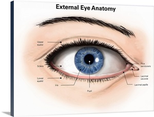
External anatomy of the human eye (with labels) Wall Art, Canvas Prints ...
The Eye - Science Quiz - Seterra Our eyes are highly specialized organs that take in the light reflected off our surroundings and transform it into electrical impulses to send to the brain. The anatomy of the eye is fascinating, and this quiz game will help you memorize the 12 parts of the eye with ease.

Human Anatomy Lab: The Urinary and Reproductive Systems
The Human Eye | Boundless Physics - Lumen Learning The human eye is the gateway to one of our five senses. The human eye is an organ that reacts with light. It allows light perception, color vision and depth perception. A normal human eye can see about 10 million different colors! There are many parts of a human eye, and that is what we are going to cover in this atom.

Label Parts of the Human Eye
Eye Diagram With Labels and detailed description A brief description of the eye along with a well-labelled diagram is given below for reference. Well-Labelled Diagram of Eye The anterior chamber of the eye is the space between the cornea and the iris and is filled with a lubricating fluid, aqueous humour. The vascular layer of the eye, known as the choroid contains the connective tissue.

WMU Psychology Department: Lisa Baker
Labeling the Human Eye Quiz - PurposeGames.com This is an online quiz called Labeling the Human Eye. There is a printable worksheet available for download here so you can take the quiz with pen and paper. This quiz has tags. Click on the tags below to find other quizzes on the same subject. Anatomy.

Label the Eye - PurposeGames
Anatomy of the Eye | Johns Hopkins Medicine The back part of the eye's interior. Pupil. The opening in the middle of the iris through which light passes to the back of the eye. Retina. The light-sensitive nerve layer that lines the inside of the back of the eye. The retina senses light and creates impulses that are sent through the optic nerve to the brain. Sclera.
31 Label The Eye Quiz - Best Labeling Ideas
Eye Anatomy: 16 Parts of the Eye & Their Functions The following are parts of the human eyes and their functions: 1. Conjunctiva The conjunctiva is the membrane covering the sclera (white portion of your eye). The conjunctiva also covers the interior of your eyelids. Conjunctivitis, often known as pink eye, occurs when this thin membrane becomes inflamed or swollen.

Label The Eye Diagram - Pensandpieces
Labelling the eye - Science Learning Hub Labelling the eye Add to collection The human eye contains structures that allow it to perceive light, movement and colour differences. In this activity, students use online or paper resources to identity and label the main parts of the human eye. By the end of this activity, students should be able to: identify the main parts of the human eye
Human Eye: Anatomy, parts and structure - Online Biology Notes
The Human Eye (Eyeball) Diagram, Parts and Pictures ... The eyeball is a round gelatinous organ that contains the actual optical apparatus. It is approximately 25 mm in diameter and sits snugly in the orbit where six muscles control its movement. The eyeball has three layers, each of which has several important structures that are essential for the sense of vision. Wall of the Eyeball
Label The Parts Of The Eye - ProProfs Quiz
Labeled Eye Diagram | Eye anatomy diagram, Eye anatomy ... This Article is the detailed account of all the major organs that are categorized under the nine regions in the abdominal cavity 1) Stomach 2) Intestines a) Small Intestine Duodenum Jejunum Ileum b) Large Intestine Ceacum Colon (Ascending, Transverse and Descending) Rectum Anal Canal 3) Liver 4) Gall bladder 5) Pancreas 6) Spleen 7) Kidneys […] S

Human Anatomy Lab: Heart Models
Eye Diagram - Labelled Diagram of Human Eye, Explanation ... The human eye is a part of the sensory nervous system. Labeled Diagram of Human Eye The eyes of all mammals consist of a non-image-forming photosensitive ganglion within the retina which receives light, adjusts the dimensions of the pupil, regulates the availability of melatonin hormones, and also entertains the body clock.

wallpapers: Horror Eye
Human eye - Wikipedia The human eye is a sensory organ, part of the sensory nervous system, that reacts to visible light and allows us to use visual information for various purposes including seeing things, keeping our balance, and maintaining circadian rhythm . The eye can be considered as a living optical device.
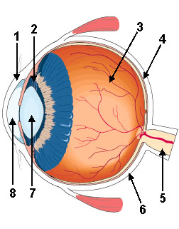
Diagram Of The Eye With Labels - ClipArt Best
Diagram Of The Eye With Labels - ClipArt Best

Free art print of Structures Of The Human Eye Labeled. Isolated ...

Amazing close-up photos of animal eyes (9 pics) | Amazing Creatures
Skeleton Clip Art at Clker.com - vector clip art online, royalty free ...
Post a Comment for "43 human eye with labels"