38 dna diagram with labels
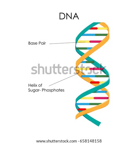
Dna Model Drawing With Labels - Solved 1 Label The Structure Of Dna Dna ... One idea would be to look up a labeled diagram of dna and label your own . If you have trouble drawing the two strands antiparallel, . You should label all the parts of the dna including the covalent and hydrogen bonds. This diagram misses out the carbon atoms in the ring for clarity. What Is DNA?- Meaning, DNA Types, Structure and Functions The DNA molecule consists of 4 nitrogen bases, namely adenine (A), thymine (T), cytosine (C) and Guanine (G), which ultimately form the structure of a nucleotide. The A and G are purines, and the C and T are pyrimidines. The two strands of DNA run in opposite directions. These strands are held together by the hydrogen bond that is present ...
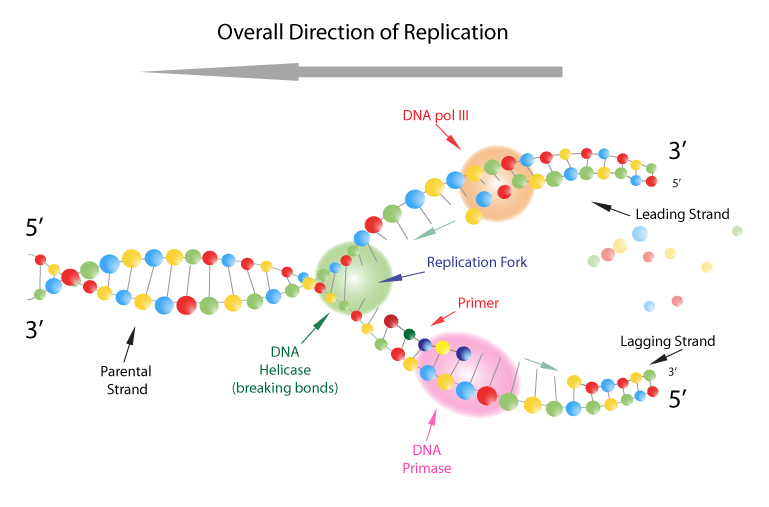
Label DNA and Replication - Google Slides Label the diagram: DNA polymerase adds nucleotides (5' to 3') Replication fork is formed. DNA polymerase attaches to the primer. Okazaki fragments bound by ligase. DNA helicase unwinds DNA. Rearrange the steps to indicate the correct order: 1. Enzyme that unwinds DNA.
Dna diagram with labels
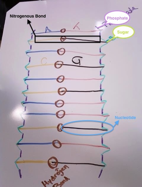
Dna Drawing With Labels / Dna Replication Steps Diagram Expii C stands for cytosine (a . The dna has twisted ladder or double helical structure. The dna molecule comprises polymers of nucleotides carrying instructions for development and growth. One idea would be to look up a labeled diagram of dna and label your own drawing. Dna Drawing With Labels / Dna Replication Steps Diagram Expii. A dna strand is made of four bases, classified with the letters a, c, t, and g. DNA Molecule Label Diagram | Quizlet Polymerase Chain Reaction (PCR) is a biochemical technique that allows scientists to take tiny samples of DNA and amplify them into large samples that can then be examined to determine the DNA sequence. (This is useful, for example, in forensic science.) The process works by mixing the sample with appropriate enzymes and then heating it until the ... The Structure of DNA The Structure of DNA This figure is a diagram of a short stretch of a DNA molecule which is unwound and flattened for clarity. The boxed area at the lower left encloses one nucleotide. Each nucleotide is itself make of three subunits: A five carbon sugar called deoxyribose (Labeled S)
Dna diagram with labels. Diagram Dna Replication Label [9YPLRS] In this diagram showing the replicationof DNA, label the following items: leading and lagging strands, Okazaki fragment, DNA polymerase, DNA ligase, helicase, primase, single-stranded binding proteins, RNA primer, replication fork, and 5' and 3' ends of the parental DNA. Okazaki concluded that DNA replication proceeds by a discontinuous mechanism. › scitable › topicpageDNA Transcription | Learn Science at Scitable - Nature The process of making a ribonucleic acid (RNA) copy of a DNA (deoxyribonucleic acid) molecule, called transcription, is necessary for all forms of life. The mechanisms involved in transcription ... Dna Replication Label Diagram [Z76A8X] DNA Replication Steps in DNA Replication • Step 2: Synthesis of DNA segments. On the diagram below, label the 5' and 3' ends of both parental DNA strands (you can make up which is which) 2. Show the complimentary base pairing that would occur in the replication of the short DNA molecule below. Dna Drawing Labeled at PaintingValley.com | Explore collection of Dna ... label art drawing labeling molecule parts idea elegant life rna worksheet information model Dna Diagram Labeled ... 1382x1382 8 1 Things You Probably ... 1563x1650 6 0 Diagram Of Dna Dvaen... 455x482 6 0 Dna Model Labeled Pa... 800x453 5 0 Here Is A Diagram Of... 600x1762 2 0 Beautiful Dna Replic... 570x243 1 0 Dna Review Sheet Dna... 630x380 1 0
dna-labeling | NEB Label: Reaction: Recommended Enzyme: DNA 5´ End Labeling: γ-32 P rATP: T4 Polynucleotide Kinase : Labeling by PCR: α-32 P dNTP, Biotin-dNTP, Fl-dNTP: Taq DNA Polymerase: DNA 3´ End Labeling: α-32 P dNTP, Biotin-dNTP, Fl-dNTP: Terminal™ Transferase. Klenow Fragment (3´ → 5´ exo-) Single Nucleotide Terminator Labeling: Fl terminator nucleotide: Therminator DNA Polymerase Animal Cell Diagram | Science Trends An animal cell diagram is a great way to learn and understand the many functions of an animal cell. The diagram, like the one above, will include labels of the major parts of an animal cell including the cell membrane, nucleus, ribosomes, mitochondria, vesicles, and cytosol. The cells of animals are the basic structural units for the wide ... DNA and RNA Probe Labeling | Radiolabeled Nucleotides Oligonucleotides can be labeled at either the 3' or the 5' end. Using polynucleotide kinase and ATP-gamma- 32 P, the 5' end is labeled. Using terminal transferase and deoxynucleotide triphosphate labeled on the alpha phosphate, the 3' end is labeled. Traditionally, the isotope of choice has been 32 P, however 35 S has been used successfully. › cells › bactcellInteractive Bacteria Cell Model - CELLS alive A specialized pilus, the sex pilus, allows the transfer of plasmid DNA from one bacterial cell to another. Pili (sing., pilus) are also called fimbriae (sing., fimbria). Flagella: The purpose of flagella (sing., flagellum) is motility. Flagella are long appendages which rotate by means of a "motor" in the cell envelope.
DNA: Structure, Forms and Functions (With Diagram) Resemblances and Differences Between B-DNA and Z-DNA: 1. Both B-DNA and Z-DNA are of double helical structure. 2. In both DNAs two strands are antiparallel. 3. G Ξ C pairing is present in both B-DNA and Z-DNA. 4. B-DNA is right-handed whereas Z-DNA is left-handed. 5. The sugar-phosphate backbone in B-DNA is regular while in Z-DNA it follows zigzag course. Mechanism of DNA Replication (explained with diagrams) | Biology In eukaryotes with large DNA molecule, there may be many initiation points (origin) of replication which finally merge with one another. 3. Unwinding of DNA molecule: The DNA double helix unwinds and uncoils into single strands of DNA by breakdown of weak hydrogen bonds. Unwinding of helix is helped by enzyme helicases. DNA Labeling: Transciption and Translation - The Biology Corner This worksheet shows a diagram of transcription and translation and asks students to label it; also includes questions about the processes. Name: _____ Review - Transcription and Translation. Label the diagram. 1. _____ 5. ... › watchEvery IB Biology drawing you NEED to know - YouTube PLEASE READ!This is every single drawing you need to know!Looking through the 2016 syllabus, this video covers 2 main types of statements- Draw- Annotate ( +...
Chromosome, Gene, DNA Diagram Label Worksheets (Differentiated) File previews. pptx, 117.44 KB. pptx, 201.71 KB. Four excellently differentiated worksheets. Engaging activity where pupils have to label a diagram containing a cell, nucleus, chromosome, gene and DNA. Very well structured and scaffolded according to ability. Excellent for visual learners. Compatible with all biology exam boards (including AQA ...
DNA Replication Labeling Diagram | Quizlet Suppose it is known that the life X of a particular compressor, in hours, has the density function $f(x)=\frac{1}{900} e^{-x/900}$, $x \gt 0$, f(x)=0, elsewhere. (a) Find the mean life of the compressor. (b) Find $E(X^2)$. (c) Find the variance and standard deviation of the random variable X. Verified answer.
› dna-color-clustering-the-leedsDNA Color Clustering: The Leeds Method for Easily Visualizing ... Aug 23, 2018 · Hi, Susan. I would definitely work with your dad’s DNA. Start by making the chart with those AncestryDNA labels a 2nd or 3rd cousins, but keep the matches under 400 shared cM. Then, see if you can label the columns as to which of your dad’s grandparents each column/cluster is related to. You can follow up here or email me at drleeds ...
› science › biologyStages of transcription: initiation, elongation & termination ... An in-depth looks at how transcription works. Initiation (promoters), elongation, and termination.
4 Ways to Use DNA Helix Diagrams in PowerPoint DNA diagram is a graphical chart resembling the shape of a double helix, a symbol of the human DNA structure that defines the characteristics of a person. This metaphor is used to illustrate an organizational culture of a corporation or smaller company, its underlying values and principles.
Methods for Labeling Nucleic Acids - Thermo Fisher Scientific A linear DNA template with this promoter sequence can be used with T7 RNA polymerase to in vitro transcribe labeled RNA probes. The +1 position indicates the first nucleotide that is incorporated into the RNA during transcription. The bases at positions +1 through +3 are critical for transcription and must be G and 2 purine bases, respectively.
Dna Structure Labeled Illustrations, Royalty-Free Vector Graphics ... Educational and medical scheme with cell, chromosome and DNA. Labeled diagram with cytosine, thymine, adenine and guanine. Teenager length compared with elderly adult. dna structure labeled stock illustrations. ... Natural product labels Natural product vector labels set. Dangerous ingredients or allergens to avoid in food, drinks and cosmetics ...
DNA Replication (With Diagram) | Molecular Biology The DNA polymerase acts on dATP, dGTP, dCTP, dTTP. These new nucleotides are added one-by-one at 3′-OH end of the growing strand. DNA polymerase needs a primer to synthesize new strand. Small RNA primer hydrogen bonds with the template. This primer provides free 3′- OH end to add new nucleotides. DNA synthesis takes place in 5′ → 3′ direction.
Genomics Genetic Testing Genetics Dna Free Clipart - Human Genome Project Png , Free Transparent ...
sdjohnston.faculty.noctrl.edu › 102 › Replication keyBio 102 Practice Problems Chromosomes and DNA Replication DNA polymerase requires a single-stranded template, a primer and nucleotides and can only add nucleotides to an existing 3’ end. In the left molecule, there are two 3’ ends that DNA polymerase could add to by reading the single-stranded segments. So DNA polymerase could add nucleotides until it reached the end of the template, as shown in
Replication Diagram Dna Label [JVASER] Search: Label Dna Replication Diagram. What is Label Dna Replication Diagram. Likes: 603. Shares: 302.
DNA Replication - Labeling.pdf - Google Drive View Details. Loading… ...
Dna diagram - Teaching resources DNA Diagram - DNA Diagram - DNA, Chromosome, Gene Diagram - DNA Replication - Diagram Simplified - DNA - DNA - DNA - DNA Review - Diagram - Planets Diagram. ... Dna labeling Labelled diagram. by Tclemmons. G7 G9 Biology. Earth's Layers Diagram Labelled diagram. by Torkelson. DNA Structure Labelled diagram. by Lrubio10.
Animal Cell Diagram with Label and Explanation: Cell ... - Collegedunia Animal cell is a typical Eukaryotic cell enclosed by a plasma membrane containing nucleus and organelles which lack cell walls, unlike all other Eukaryotic cells. The typical cell ranges in size between 1-100 micrometers. The lack of cell walls enabled the animal cells to develop a greater diversity of cell types.
Dna Model Drawing With Labels : Dna Model Biology Junction One idea would be to look up a labeled diagram of dna and label your own . C stands for cytosine (a . Genes are the genetic component that is found compressed in the nucleus of the cell. The dna has twisted ladder or double helical structure. A stands for adenine (a purine); Dna is a double stranded structure, and . The above structure is a .
DNA function & structure (with diagram) (article) | Khan Academy Chromosomal DNA consists of two DNA polymers that make up a 3-dimensional (3D) structure called a double helix. In a double helix structure, the strands of DNA run antiparallel, meaning the 5' end of one DNA strand is parallel with the 3' end of the other DNA strand.

Selina Concise Class 10 Biology Chapter 2 Structure of Chromosomes, Cell Cycle and Cell Division ...
Dna labeling - Labelled diagram - Wordwall Dna labeling - Labelled diagram Sugar (Deoxyribose), phosphate , nitrogen base Adenine, nitrogen base Guanine, Hydrogen bond, nitrogen base cytosine, nitrogen base thymine, backbone. Dna labeling Share by Tclemmons G7 G9 Biology Like Edit Content More Log in required Theme Log in required Options Switch template Interactives






Post a Comment for "38 dna diagram with labels"