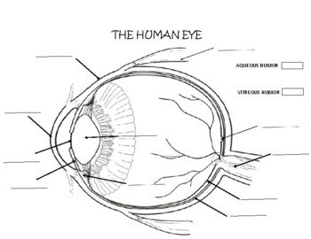
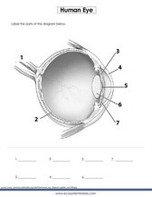
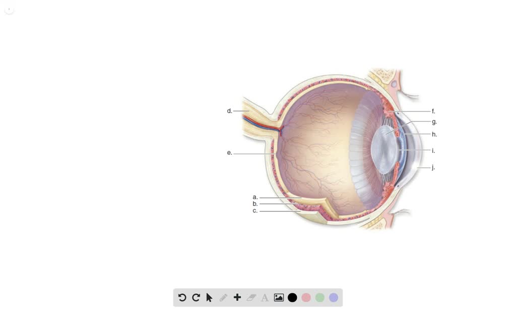
43 the human eye without labels
20 Facts About the Amazing Eye - Discovery Eye Foundation Here are a few facts you may enjoy: 1. Eyes began to develop 550 million years ago. The simplest eyes were patches of photoreceptor protein in single-celled animals. 2. Your eyes start to develop two weeks after you are conceived. 3. The entire length of all the eyelashes shed by a human in their life is over 98 feet with each eye lash having a ... human eye diagram with labels Human Skeleton Blank Clip Art at Clker.com - vector clip art online. 8 Pics about Human Skeleton Blank Clip Art at Clker.com - vector clip art online : Human Eye Diagrams with the Unlabeled, picture front of the eye without labels clipart - Clipground and also Male Reproductive System | Free Images at Clker.com - vector clip art.
human eye diagram with labelling Eye human diagram label scholastic ks2 children related. Eye blank diagram fill clipart. Diagram of the human eye - primary ks2 teaching resource ... eye diagram anatomy labeled labels without label human science parts enchantedlearning labelled eyeball eyes grade clipart inside learn worksheets teaching. Eye Diagram: Label Quiz

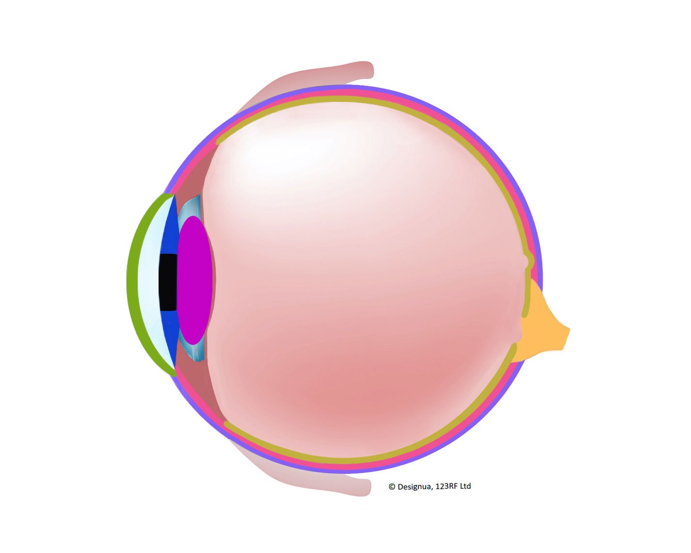
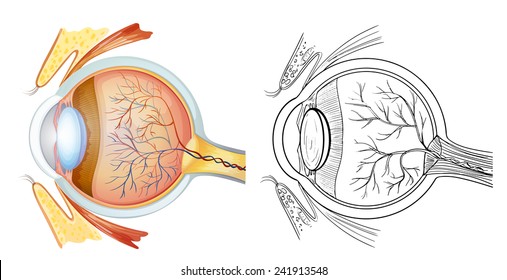
The human eye without labels
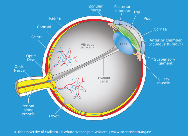
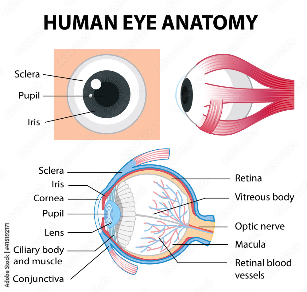
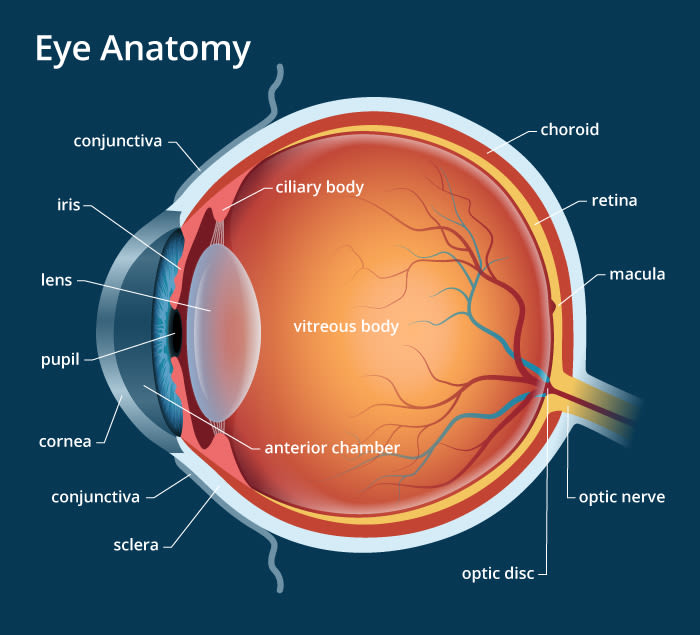
Normal chest MDCT with anatomic labels | e-Anatomy - e-Anatomy … 10.3.2022 · Pocket Atlas of Human Anatomy: 5th edition - W. Dauber, Founded by Heinz Fene Anatomical variants and notes from the author about the anatomical labeling of the thorax CT: In the lower lobe of the left lung, there is an inconstant subsuperior pulmonary segment that is seen in approximately 30% of individuals, located between the superior and basal segments of the … Interaction Design vs UX: What's the Difference? - Adobe XD 16.10.2019 · Interaction designers apply physiological principles to the design of products. The goal of this process is to reduce human error, increase productivity, and enhance the safety of interaction. Interaction designers often use a predictive model of human movement, also known as Fitts’s law, when they design interactions. The Human Eye - Diagram, Parts, Working, Function and Work of The Lens Sclera: The sclera is the protective outer layer, a strong white coating that protects the eyes (white part of the eye). Cornea: The cornea is the sclera's translucent front part. The cornea allows light to flow through and into the eye. Iris: The iris is a black muscular tissue and ring-like structure behind the cornea. The eye's colour is determined by the colour of the iris.
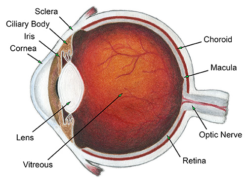
The human eye without labels. Category:Human eyes - Wikimedia Commons Black eyes by megamoto85 (cropped).jpg 925 × 673; 148 KB Blue Eyed Girl - Flickr - rcstanley.jpg 529 × 622; 49 KB Blue-Green Eye miosis.jpg 917 × 688; 451 KB apps.apple.com › us › appThe Human Body by Tinybop 4+ - App Store + Feed the body, make it run and breathe, assemble and pull apart a skeleton, see how the eye sees, watch sound vibrations travel through the ear canal, and more. + Learn new vocabulary with text labels in 50+ languages. + Create a dashboard to change languages, delete accounts, and support your kids’ learning. Anatomy of the Eye | Kellogg Eye Center | Michigan Medicine Layer containing blood vessels that lines the back of the eye and is located between the retina (the inner light-sensitive layer) and the sclera (the outer white eye wall). Ciliary Body. Structure containing muscle and is located behind the iris, which focuses the lens. Cornea. The clear front window of the eye which transmits and focuses (i.e ... Human eye - Wikipedia The human eye is a sensory organ, part of the sensory nervous system, that reacts to visible light and allows us to use visual information for various purposes including seeing things, keeping our balance, and maintaining circadian rhythm . The eye can be considered as a living optical device.
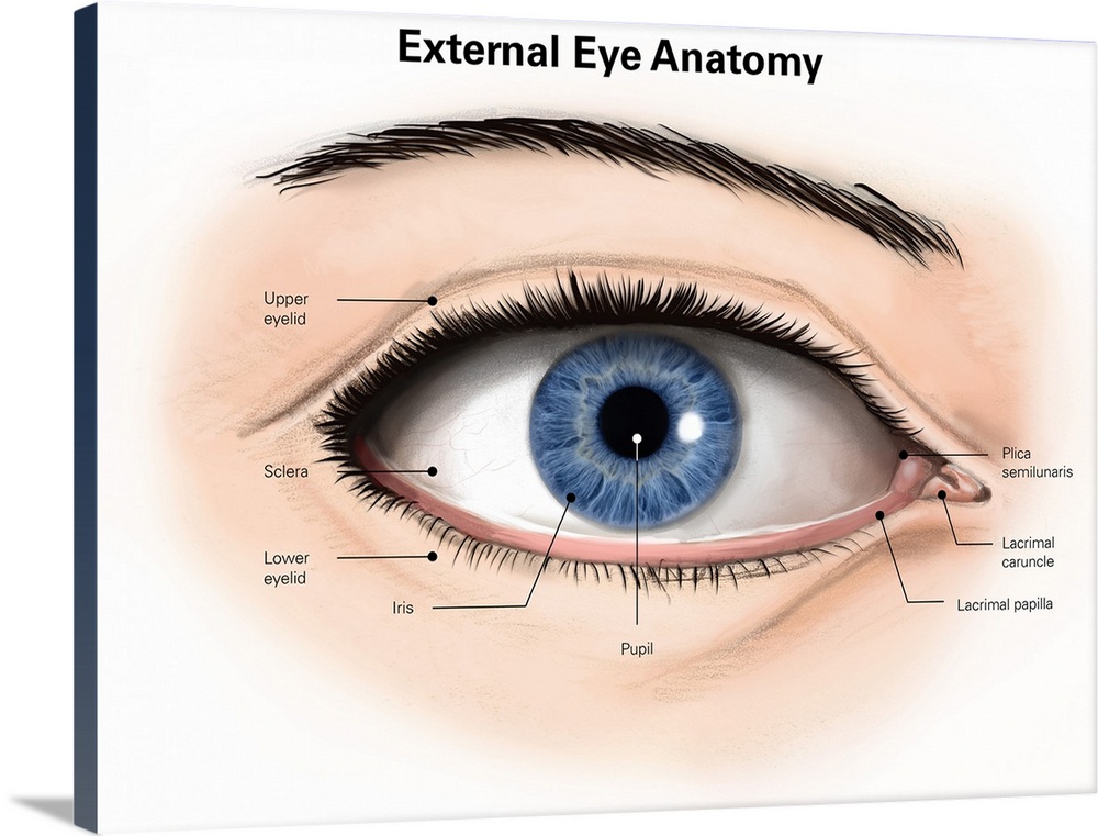
Human Eye - Definition, Structure, Function, Parts, Diagram - BYJUS Structure of Human Eye. A human eye is roughly 2.3 cm in diameter and is almost a spherical ball filled with some fluid. It consists of the following parts: Sclera: It is the outer covering, a protective tough white layer called the sclera (white part of the eye). Cornea: The front transparent part of the sclera is called the cornea. Eye Anatomy: Parts of the Eye and How We See Behind the anterior chamber is the eye's iris (the colored part of the eye) and the dark hole in the middle called the pupil. Muscles in the iris dilate (widen) or constrict (narrow) the pupil to control the amount of light reaching the back of the eye. Directly behind the pupil sits the lens. The lens focuses light toward the back of the eye. Label Parts of the Human Eye - University of Dayton Parts of the Eye. Select the correct label for each part of the eye. The image is taken from above the left eye. Click on the Score button to see how you did. Incorrect answers will be marked in red. ... › Can-C-Drops-Milliliter-LiquidAmazon.com: Can-C Eye Drops 5ml Liquid (2 in 1 Pack) Can C ... This item: Can-C Eye Drops 5ml Liquid (2 in 1 Pack) Can C Cataract Eye Drops N-Acetylcarnosine, Human and Animal Eye, Cataract Eye Drops for Dog - Gift Set with Boxiti Wipe $39.00 $ 39 . 00 ($229.41/Fl Oz)
Amazon.com: Can-C Eye Drops 5ml Liquid (2 in 1 Pack) Can C Cataract Eye ... After one week of using Can -C my vision became clearer and more crisp. After 3 weeks, I have been able to see my phone and iPad without my reading glasses, I have been driving without my glasses, and I have been watching TV without my glasses. I can read the Guide on my TV to see what's on the TV. And, my dry eye has improved as well. human eye | Definition, Anatomy, Diagram, Function, & Facts The protrusion of the eyeballs—proptosis—in exophthalmic goitre is caused by the collection of fluid in the orbital fatty tissue. The eyelids eyelid It is vitally important that the front surface of the eyeball, the cornea, remain moist. Structure and Function of the Human Eye - ThoughtCo Key Takeaways: The Human Eye and Vision The main parts of the human eye are the cornea, iris, pupil, aqueous humor, lens, vitreous humor, retina, and optic nerve. Light enters the eye by passing through the transparent cornea and aqueous humor. The iris controls the size of the pupil, which is the opening that allows light to enter the lens. File:Diagram of human eye without labels.svg - Wikimedia Size of this PNG preview of this SVG file: 410 × 430 pixels. Other resolutions: 229 × 240 pixels | 458 × 480 pixels | 732 × 768 pixels | 976 × 1,024 pixels | 1,953 × 2,048 pixels. Original file (SVG file, nominally 410 × 430 pixels, file size: 277 KB) File information. Structured data.
Anatomy of the eye: Quizzes and diagrams | Kenhub Here you can see all of the main structures in this area. Spend some time reviewing the name and location of each one, then try to label the eye yourself - without peeking! - using the eye diagram (blank) below. Unlabeled diagram of the eye Click below to download our free unlabeled diagram of the eye.
Guidelines for Safe Work Practices in Human and Animal … 6.1.2012 · All functions of the human and animal diagnostic laboratory — microbiology, chemistry, ... (44), splash in eye (19), and bruises and cuts (45) as the most frequent ... Without good housekeeping, any other preventive measures (e.g., installation of sophisticated flooring, specialty footwear, or training on techniques of ...
Quiz: Label The Parts Of The Eye - ProProfs Quiz Take up this quiz and find out how much did you get to understand about the human eye? All the very best to you! Questions and Answers 1. A is pointing to what part of the eye? A. Cornea B. Optic Nerve C. Iris D. Pupil E. Sclera 2. B is pointing to what part of the eye? A. Optic Nerve B. Lens C. Retina D. Pupil E. Iris 3.
› health › can-you-see-bed-bugsCan You See Bedbugs: Size, Identification, Removal, Prevention Sep 24, 2019 · Adult bedbugs are visible to the human eye. They're about the size of an apple seed and reddish-brown in color. Learn how to identify these unwelcome critters, how to get rid of them, and how to ...
en.wikipedia.org › wiki › Human_penisHuman penis - Wikipedia The human penis is an external male intromittent organ that additionally serves as the urinary duct. The main parts are the root (radix); the body (corpus); and the epithelium of the penis including the shaft skin and the foreskin (prepuce) covering the glans penis .
Labeling Quizlet Eye [T0VN93] Interactive Nutrition Facts Label Official Written Warning Template Can you locate the parts of the human eye? by smac17 Plays Quiz Updated Nov 13, 2017 Lens: The lens is a clear part of the eye behind the iris that helps Retina -The retina is the area at the back of the eye that receives the refined, visual message from the front of the eye ...
PDF Eye Anatomy Handout - National Institutes of Health of light entering the eye. Lens: The lens is a clear part of the eye behind the iris that helps to focus light, or an image, on the retina. Macula: The macula is the small, sensitive area of the retina that gives central vision. It is located in the center of the retina. Optic nerve: The optic nerve is the largest sensory nerve of the eye.
Eye Anatomy: A Closer Look At the Parts of the Eye - All About Vision In a number of ways, the human eye works much like a digital camera: Light is focused primarily by the cornea — the clear front surface of the eye, which acts like a camera lens. The iris of the eye functions like the diaphragm of a camera, controlling the amount of light reaching the back of the eye by automatically adjusting the size of the ...
xd.adobe.com › ideas › principlesInteraction Design vs UX: What's the Difference? - Ideas Oct 16, 2019 · Words encompass text, which helps convey the right amount of information to users. Words, especially those used in interactions, like button labels, should be meaningful and simple to understand. They should communicate information to users without overwhelming them with too many details. 2D: Visual representations.
PDF Parts of the Eye - National Institutes of Health Eye Diagram Handout Author: National Eye Health Education Program of the National Eye Institute, National Institutes of Health Subject: Handout illustrating parts of the eye Keywords: parts of the eye, eye diagram, vitreous gel, iris, cornea, pupil, lens, optic nerve, macula, retina Created Date: 12/16/2011 12:39:09 PM
Lens of the Eye - All About Vision The lens of the eye, also called the crystalline lens, is an important part of the eye's anatomy that allows the eye to focus on objects at varying distances. It is located behind the iris and in front of the vitreous body. In its natural state, the lens looks like an elongated sphere — a shape known as ellipsoid — that resembles a ...
Human penis - Wikipedia The human penis is an external male intromittent organ that additionally serves as the urinary duct.The main parts are the root (radix); the body (corpus); and the epithelium of the penis including the shaft skin and the foreskin (prepuce) covering the glans penis.The body of the penis is made up of three columns of tissue: two corpora cavernosa on the dorsal side and corpus …
The Eyes (Human Anatomy): Diagram, Optic Nerve, Iris, Cornea ... - WebMD The weaker eye, which may or may not wander, is called the "lazy eye." Astigmatism: A problem with the curve of your cornea. If you have it, your eye can't focus light onto the retina the way it...
Eye Defects And Corrections in Humans; Reasons, Effect and Solution The human eye is the most important organ in the body. The eye serves as the king as nothing can be done easily without the eyes. Eyes are used mainly for sight. This delicate organ needs proper care and attention because its loss will throw a being into total darkness. Some people are born without sight while some develop sight defects over time.
Amazon.com: Eye Magic Premium Instant Eyelid Lift (S/M Kit … Our Instant Eye Lift is specifically designed to gently lift and define droopy upper eyelids for a more youthful appearance. It’s like getting a new, youthful, layer of eyelid skin. You can apply our “eye lifts” with (or without) makeup in the morning and remove before bed. How and when you use our eye lift product is up to you.
Human Eye Explorer Even microscopic structures, usually not visible for the human eye, can be explored: see inside the retina or cornea and discover its layers and cells from all perspectives. Additional functions like the presentation editor, the media library or labeling and color tools, make the Human Eye Explorer a unique software.
The Human Body by Tinybop 4+ - App Store + Feed the body, make it run and breathe, assemble and pull apart a skeleton, see how the eye sees, watch sound vibrations travel through the ear canal, and more. + Learn new vocabulary with text labels in 50+ languages. + Create a dashboard to change languages, delete accounts, and support your kids’ learning.
Eye Anatomy: 16 Parts of the Eye & Their Functions - Vision Center The lens of the eye (or crystalline lens) is the transparent lentil-shaped structure inside your eye. This is the natural lens. It is located behind the iris and to the front of the vitreous humor (vitreous body). The vitreous humor is a clear, colorless, gelatinous mass that fills the gap between the lens and the retina in the eye.
What Does the Eye Look Like? - Diagram of the Eye | Harvard Eye Associates Vitreous Gel: A thick, transparent liquid that fills the center of the eye. It is mostly water and gives the eye its form and shape. Our eyes are vital for seeing the world around us. Keep them healthy by maintaining regular vision exams. Contact Harvard Eye Associates at 949-951-2020 or harvardeye.com to schedule an appointment today.
Can You See Bedbugs: Size, Identification, Removal, Prevention 24.9.2019 · Adult bedbugs are visible to the human eye. They're about the size of an apple seed and reddish-brown in color. Learn how to identify these unwelcome critters, how to get rid of them, and how to ...
› en › e-AnatomyNormal chest MDCT with anatomic labels | e-Anatomy - IMAIOS Mar 10, 2022 · Pocket Atlas of Human Anatomy: 5th edition - W. Dauber, Founded by Heinz Fene Anatomical variants and notes from the author about the anatomical labeling of the thorax CT: In the lower lobe of the left lung, there is an inconstant subsuperior pulmonary segment that is seen in approximately 30% of individuals, located between the superior and ...
› mmwr › previewGuidelines for Safe Work Practices in Human and Animal ... Jan 06, 2012 · In clinical chemistry laboratories, data from 17 New York hospitals listed needle puncture (103 cases), acid or alkali spills , glass cuts (44), splash in eye (19), and bruises and cuts (45) as the most frequent exposures (21). Needle puncture, glass cuts, splash in eye, and bruises and cuts have the highest potential for infection from microbes.
60,892 Human eye anatomy Images, Stock Photos & Vectors - Shutterstock Find Human eye anatomy stock images in HD and millions of other royalty-free stock photos, illustrations and vectors in the Shutterstock collection. Thousands of new, high-quality pictures added every day.
Eye Diagram With Labels and detailed description - BYJUS Iris is the coloured part of the eye and controls the amount of light entering the eye by regulating the size of the pupil. The lens is located just behind the iris. Its function is to focus the light on the retina. The optic nerve transmits electrical signals from the retina to the brain. Pupil is the opening at the centre of the iris.
label the ear worksheet Picture Front Of The Eye Without Labels Clipart 20 Free Cliparts clipground.com eye human diagram worksheet eyeball learning layers without anatomy labels parts eyes worksheets clipart science structure grade clipground body structures 14 Best Images Of Ear Hearing Worksheets - Listening Ear Craft Template
Structure of the Human Eye - Health Jade The eye is a hollow, spherical structure about 2.5 centimeters in diameter. Its wall has three distinct layers—an outer (fibrous) layer, a middle (vascular) layer, and an inner (nervous) layer. The spaces within the eye are filled with fluids that help maintain its shape. Figure 6. Structure of the human eye Outer Layer of the Eye
The Human Eye - Diagram, Parts, Working, Function and Work of The Lens Sclera: The sclera is the protective outer layer, a strong white coating that protects the eyes (white part of the eye). Cornea: The cornea is the sclera's translucent front part. The cornea allows light to flow through and into the eye. Iris: The iris is a black muscular tissue and ring-like structure behind the cornea. The eye's colour is determined by the colour of the iris.
Interaction Design vs UX: What's the Difference? - Adobe XD 16.10.2019 · Interaction designers apply physiological principles to the design of products. The goal of this process is to reduce human error, increase productivity, and enhance the safety of interaction. Interaction designers often use a predictive model of human movement, also known as Fitts’s law, when they design interactions.
Normal chest MDCT with anatomic labels | e-Anatomy - e-Anatomy … 10.3.2022 · Pocket Atlas of Human Anatomy: 5th edition - W. Dauber, Founded by Heinz Fene Anatomical variants and notes from the author about the anatomical labeling of the thorax CT: In the lower lobe of the left lung, there is an inconstant subsuperior pulmonary segment that is seen in approximately 30% of individuals, located between the superior and basal segments of the …











:max_bytes(150000):strip_icc()/GettyImages-695204442-b9320f82932c49bcac765167b95f4af6.jpg)








Post a Comment for "43 the human eye without labels"